- Home
- Health Science
- Tendon Trouble
Tendon Trouble
NIH NEWS IN HEALTH
NIH News in Health · Source article
Tendons connect muscles to bones. Without them, your muscles couldn’t move your body. When you injure a tendon, it can affect your everyday activities.
“No matter how strong your muscle is, you’re not going to be able to have the functionality and the stability that you need if a tendon is impaired,” says Dr. Nelly Andarawis-Puri, a bioengineering researcher at Cornell University.
Tendon troubles can arise from The heat, swelling and redness caused by the body’s protective response to injury or infection. inflammation, a rupture, or breakdown (degeneration) of the tendon tissue. These conditions are called tendinopathies. Symptoms include swelling, pain, stiffness, and weakness.
Problems can stem from a sudden, awkward movement. But they’re usually a result of overuse. Overuse injuries are common in athletes. But daily activities that require repetitive movements can put anyone at risk. For instance, repeated exercises that require jumping may cause knee problems. Typing all day at work may lead to pain in your hands.
Tendon injuries can lead to long-term (chronic) issues if they don’t fully heal. And they often don’t. That’s because our bodies’ ability to repair tendon tissue declines with age.
Researchers are trying to unravel why tendons often don’t heal properly and what’s needed to repair them.
Breaking the Cycle
“The most common type of tendon injury is an overuse, or wear-and-tear, injury,” explains Andarawis-Puri. “It’s not necessarily painful once it starts and can be silent for a long time. Essentially, you predispose yourself to more and more accumulation of damage. Then, eventually you have degeneration.”
Getting treatment early can help keep tendon problems from getting worse. Your doctor may recommend icing the area, pain relievers, and resting the tendon.
These treatments don’t help repair your tendon. “But they can make you less miserable and manage the pain and the discomfort,” Andarawis-Puri says.
Physical therapy is often recommended for treatment as well. But there is a fine balance between exercise that promotes healing or causes damage. “That’s tricky,” Andarawis-Puri says, “because that’s different for every person.”
Her team is studying the biology of tendons to better understand how and when exercise promotes healing.
Tendons are made up of cells called tenocytes and bundles of a protein called collagen. These bundles form long helical chains.
“These helices look almost like springs—that allows them to coil and uncoil,” says Dr. Adam Abraham, an expert on chronic tendon disease at the University of Michigan. Coiling helps the fibers absorb the force from your body movements.
“A tendon is a unique connective tissue that’s highly ordered and made primarily of collagen. That’s what gives it its strength,” explains Dr. Jenna Galloway, a regenerative medicine expert at Massachusetts General Hospital and Harvard Medical School. “When you do too much movement, you can damage some of those collagen fibers. Normally, we can repair a little bit of damage. But if you do this over and over again, you change the properties of the tendon.”
When collagen fibers are injured, they look kinked under a microscope, says Andarawis-Puri. Her team has shown in animals that exercising too soon after an injury worsens that kinking. They’ve also found that waiting two weeks before letting the animals exercise gives time for the coils to straighten out.
“Our research shows that with the right timing and conditions, movement can actually trigger the tendon to repair,” Andarawis-Puri says.
Her team hopes to find ways to easily tell when exercise will help rather than hurt.
Reversing Chronic Conditions
The highly organized nature of tendons is key for them to work properly.
“But tendons are a rather lazy organ,” Abraham explains. “They are slow to adapt, if they adapt at all.”
Abraham’s team is trying to understand the changes tendons undergo from chronic injuries. They’ve developed a system that allows them to grow 3D tendon-like structures using cells from patients’ tendons.
“We’re developing a system that basically allows us to make microtendons,” Abraham explains. “The goal is to grow hundreds to thousands of these from a single person. That may sound very large, but they’re the size of a human hair.”
This 3D system allows the researchers to expose the micro-tendons to different conditions. Then, they can look at how the microtendons respond. They’re comparing microtendons grown with cells taken from people who have healthy tendon tissue to those with chronic tendinopathies.
“We can stress out healthy microtendons and get them to behave like those from patients with chronic injuries,” says Abraham. “The cells that come from patients with a chronic condition are already stressed. They become locked in this stressed state.”
His team is trying to restore chronically injured cells to a healthy state. “We’re hoping with this 3D system, we will be able to synthesize many hundreds of different environments at the same time. So we can screen what might be a possible fix,” he explains.
Abraham’s team is also testing an injectable hydrogel to mimic a healthy environment. They hope to deliver it to injured tissue to restore an organized tendon structure.
Coaxing Repair
Unlike people, some animals can fully repair tendon tissue as adults. For instance, zebrafish completely regenerate tendons throughout their lives. Galloway hopes to learn from them.
Her group has shown that tenocytes can repair fully torn tendon tissue in zebrafish. Her recent study showed that the cells bridge fully severed tissue back together.
People and other mammals have tenocytes, too. But after a tendon is injured in mammals, the tissue usually doesn’t recover. Instead, a scar forms and disrupts the carefully crafted collagen structure.
There’s something different about how mammals respond to a fully torn tendon injury, Galloway says.
“The cells in the zebrafish can respond to an injury to regenerate the tissue, but the cells in mammals cannot,” she adds.
Her team is comparing differences between fish and mammals after a tendon injury. And they’re testing potential compounds in injured tissue in mammals. They hope they can make the process of healing go better for them, too.
Scientists are still trying to fully understand tendon biology. In the meantime, it’s important to protect your tendons (see the Wise Choices box) and get injuries treated early.
Further reading and research references
- 7 Tips: Nutritional Approaches for Musculoskeletal Inflammation
- Sports Injuries–Types, Symptoms, & Risk Factors
- Nutritional Approaches for Musculoskeletal Pain and Inflammation
- Histological and immunohistochemical guide to tendon tissue.
- Glycosaminoglycans modulate microscale mechanics and viscoelasticity in fatigue injured tendons.
- Endogenous tenocyte activation underlies the regenerative capacity of the adult zebrafish tendon.
- Engineered microenvironmental cues from fiber-reinforced hydrogel composites drive tenogenesis and aligned collagen deposition.
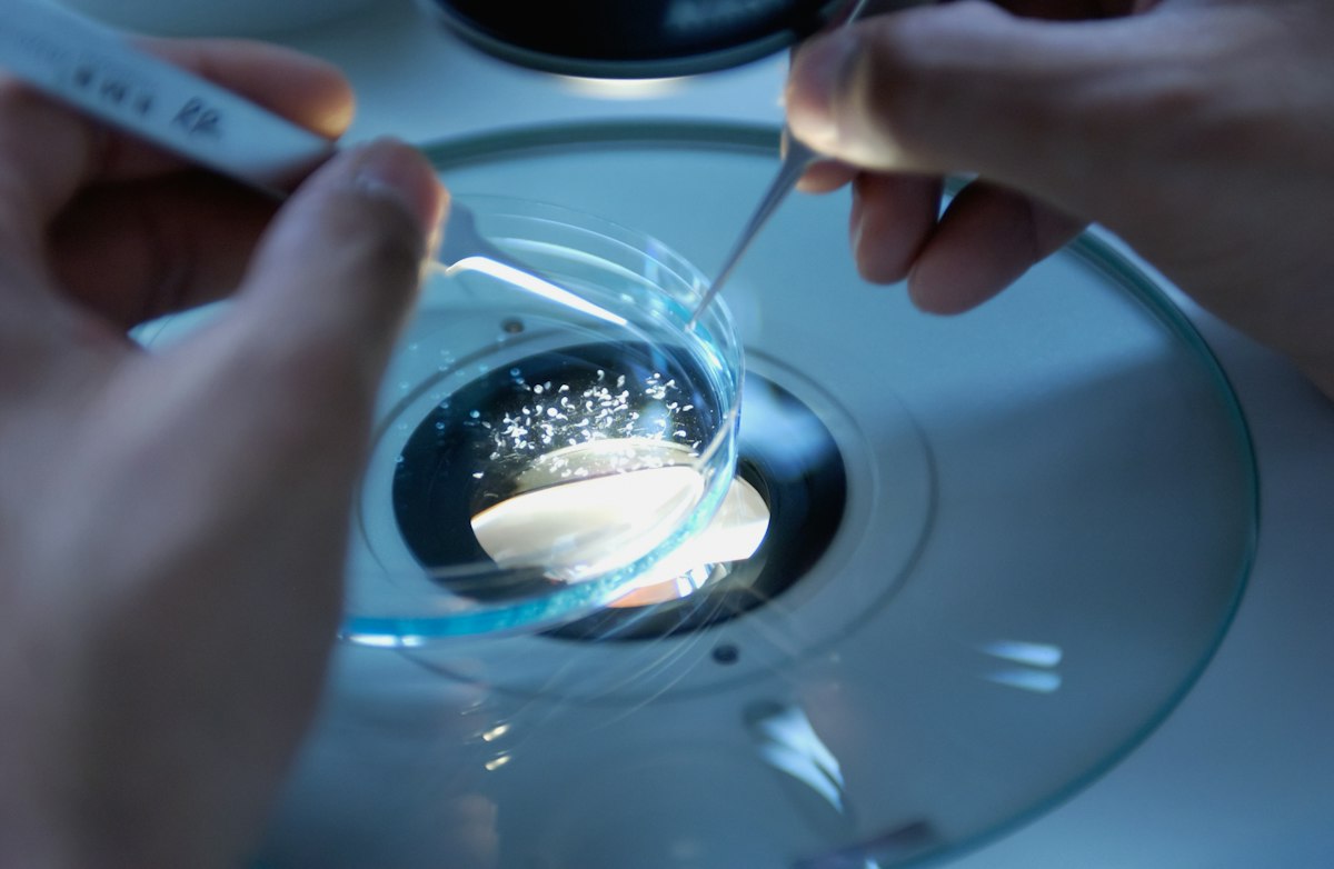
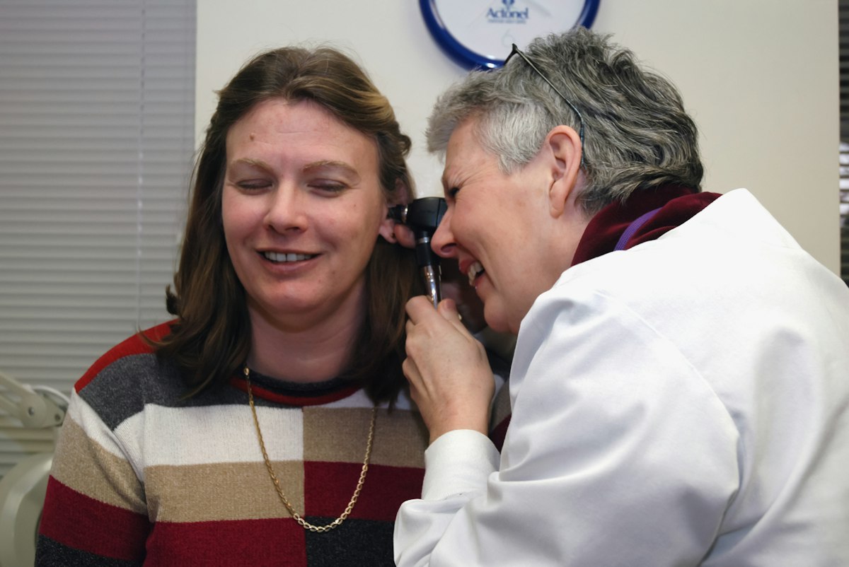
Illustrative archival photograph: National Cancer Institute / Unsplash, used under the Unsplash License. The people or equipment shown are not identified as participants in the research or interviews described here.
Health Science
View more
NIH News in Health · Source article Health Capsule Most of us have some decline in our memory and learning abilities as we age. But certain older adults can remember past events better than most people 25 to 30 years younger. Researchers recently found that exceptional recall and memory is linked to the ability to create new brain cells. An NIH-funded research team analyzed more than 350,000 individual brain cells from younger and older adults, including cognitive SuperAgers. Cognitive SuperAgers are people at least 80 years old with memory test scores as good as or better than people in their 50s and 60s. The cells were from donated postmortem brains. The team looked for newly formed brain cells. They found that the molecular make up of new brain cells differed depending on the donor’s cognitive abilities. SuperAgers’ brain cells had a unique molecular make-up. SuperAgers also had more newly created brain cells than other groups. Certain cell processes in the SuperAgers’ brains appeared to be better maintained, too. These processes serve important functions. Some are involved in communication between brain cells. Others help with cell survival. “Determining why some brains age more healthily than others can help researchers make therapeutics that will boost memory in aging and prevent Alzheimer’s disease and related dementias,” says Dr. Orly Lazarov at the University of Illinois, Chicago, who co-led the study. Source: NIH News in Health. Originally published June 2026. Republished by Graymedy on 11 October 2026. This is a sourced NIH article, not a Graymedy interview or original study. Read the original article and its research references. NIH News in Health permits republication of its articles and illustrations with acknowledgement. Research findings retain their original study context. Further reading and research references Dealing with Dementia: When Thinking and Behavior Decline What is Dementia? Symptoms, Types, and Diagnosis Alzheimer's Disease & Related Dementias Human hippocampal neurogenesis in adulthood, ageing and Alzheimer's disease Illustrative archival photograph: National Cancer Institute / Unsplash, used under the Unsplash License. The people or equipment shown are not identified as participants in the research or interviews described here.
DASH-Style Diet Helps Control Blood Sugar
Tendon Trouble
Neighborhood Design Impacts Walking Levels
NIH News in Health · Source article Health Capsule Arsenic is a naturally occurring element. It’s found in soil, water, and air. Long-term exposure can cause health problems. Millions of people rely on water sources with unsafe levels of arsenic. A new study showed that reducing arsenic exposure can lower the chances of death from cancer and heart disease. The study tracked adults living in Bangladesh. Researchers measured the levels of arsenic in participants’ drinking water and urine. They checked levels multiple times over 20 years. During those years, a mitigation program labeled unsafe wells. The government also installed deeper wells with less arsenic. The scientists tracked deaths from cancer and heart disease over the two decades. Participants’ arsenic levels dropped by over one-half on average. Their chances of dying from cancer and heart disease also changed. Those whose arsenic levels dropped were less likely to die. People whose exposures rose over time had a higher risk of death. Some participants’ arsenic levels started out high but ended low. They were half as likely to die than people whose levels remained high. “Our research suggests that improved access to low-arsenic water, periodic testing, and clear signage to mark safer water are useful tools to help improve long-term health around the world,” says Dr. Yu Chen at NYU Langone Health. Source: NIH News in Health. Originally published February 2026. Republished by Graymedy on 11 October 2026. This is a sourced NIH article, not a Graymedy interview or original study. Read the original article and its research references. NIH News in Health permits republication of its articles and illustrations with acknowledgement. Research findings retain their original study context. Further reading and research references Arsenic Factsheet Arsenic and Drinking Water (USGS) ToxFAQs™ for Arsenic (CDC) Arsenic exposure reduction and chronic disease mortality. Illustrative archival photograph: National Cancer Institute / Unsplash, used under the Unsplash License. The people or equipment shown are not identified as participants in the research or interviews described here.
The World Inside Your Mouth
Protect Against Air Pollution

Bacteria Linked to Kidney Stone Formation
NIH News in Health · Source article Burns can affect anyone. The consequences can range from mild skin damage to permanent scarring or even death. That’s why it’s important for everyone to know how to prevent burns. Most burn injuries occur at home. Kitchen fires or other household accidents are common causes. For children, scalds are the leading cause of burn injury. These are caused by steam or boiling water. Treatment depends on how serious the burn is. A first-degree burn affects only the outer layer of skin, called the epidermis. Sunburns are one example. You can usually treat them at home by cleaning the area and applying lotion to keep it moist. Second-degree burns are deeper. They affect the second layer of skin, called the dermis. These burns may cause scars or change how the skin feels. Some can be treated with wound care only. This includes washing the wound and covering it with a bandage. Deep second-degree burns may require a surgery called skin grafting. This is where healthy skin is removed from one area of the body and used to cover the damaged area. Third-degree burns are even deeper. They damage the entire skin down to the fat and nerves beneath the dermis. Fourth-degree burn cases are the most serious. Muscles, tendons, and bones can also be affected. These burns always require medical care. Treatment often includes skin grafting surgery. Doctors determine how serious a burn is through visual examination. “Burn injuries often worsen after the initial injury, so it can take up to a week to get an accurate assessment,” says Dr. Angela Gibson, a surgeon who specializes in burn care at the University of Wisconsin. Gibson and her team are studying an imaging technology to help determine how deep a burn is. The tool uses fluorescent dyes to see into the skin. If successful, the tool may help doctors more accurately predict burn depth. This could allow patients to get the right care sooner. It could also help doctors identify healthy tissue, so they remove only damaged tissue. “We want to limit the amount of healthy skin we cut away,” Gibson explains. “If you cut away tissue, you must replace it with the patient’s skin. In those cases, we’d like to keep the area where the skin was taken for the skin graft, or donor site, as small as we can.” Gibson’s lab is also studying bandages that generate tiny electrical signals. These signals tell cells to grow and move to the injured area. This helps new skin form and supports the growth of new blood vessels. The bandages copy the body’s natural electrical activity, which helps burn injuries heal faster. Traditional electrical stimulation therapy needs an external power source. But the bandages use the body’s movements. Movement creates electricity to power the bandages. This technology is being studied for multiple uses, including wound healing. But it’s still in the early stages and needs more research. Scientists continue to study new treatment options. But Gibson says prevention is the best defense. She recommends burn prevention education for everyone. Learn ways to help prevent burns at home. See the Wise Choices box for tips. “A burn injury can happen to anyone,” Gibson says. “Prevention is just as important as knowing how to care for burns.” Source: NIH News in Health. Originally published May 2026. Republished by Graymedy on 11 October 2026. This is a sourced NIH article, not a Graymedy interview or original study. Read the original article and its research references. NIH News in Health permits republication of its articles and illustrations with acknowledgement. Research findings retain their original study context. Further reading and research references Learn About Burn Care Burn Prevention, First Aid and Calling 911(FEMA) Protect the ones you love (CDC) References Indocyanine green and protoporphyrin IX fluorescence imaging of inflammation, hypoxia, and necrosis of burns Burn progression in human skin—A review of current knowledge and opportunities for future research Recent advances of nanogenerator technology for cardiovascular sensing and monitoring Illustrative archival photograph: National Cancer Institute / Unsplash, used under the Unsplash License. The people or equipment shown are not identified as participants in the research or interviews described here.
Certain Proteins May Help Prevent Food Allergies
Understanding Huntington's